Monozygotic twins – more than equal

In an issue of Genome Biology in January 2018, Van Baak and colleagues analyzed the epigenome of monozygotic versus dizygotic twins. As you can see from the figure which was copied from the OpenAccess journal, dizygotic twins expose divergent methylation patterns in several genes (in red). Most remarkably is this for the DUSP22 gene which codes for a “dual specificity phosphatase”. On the other hand, monozygotic twins (in blue) showed almost no divergence, methylation rate ratio were very close to one, which would be on the diagonal line.

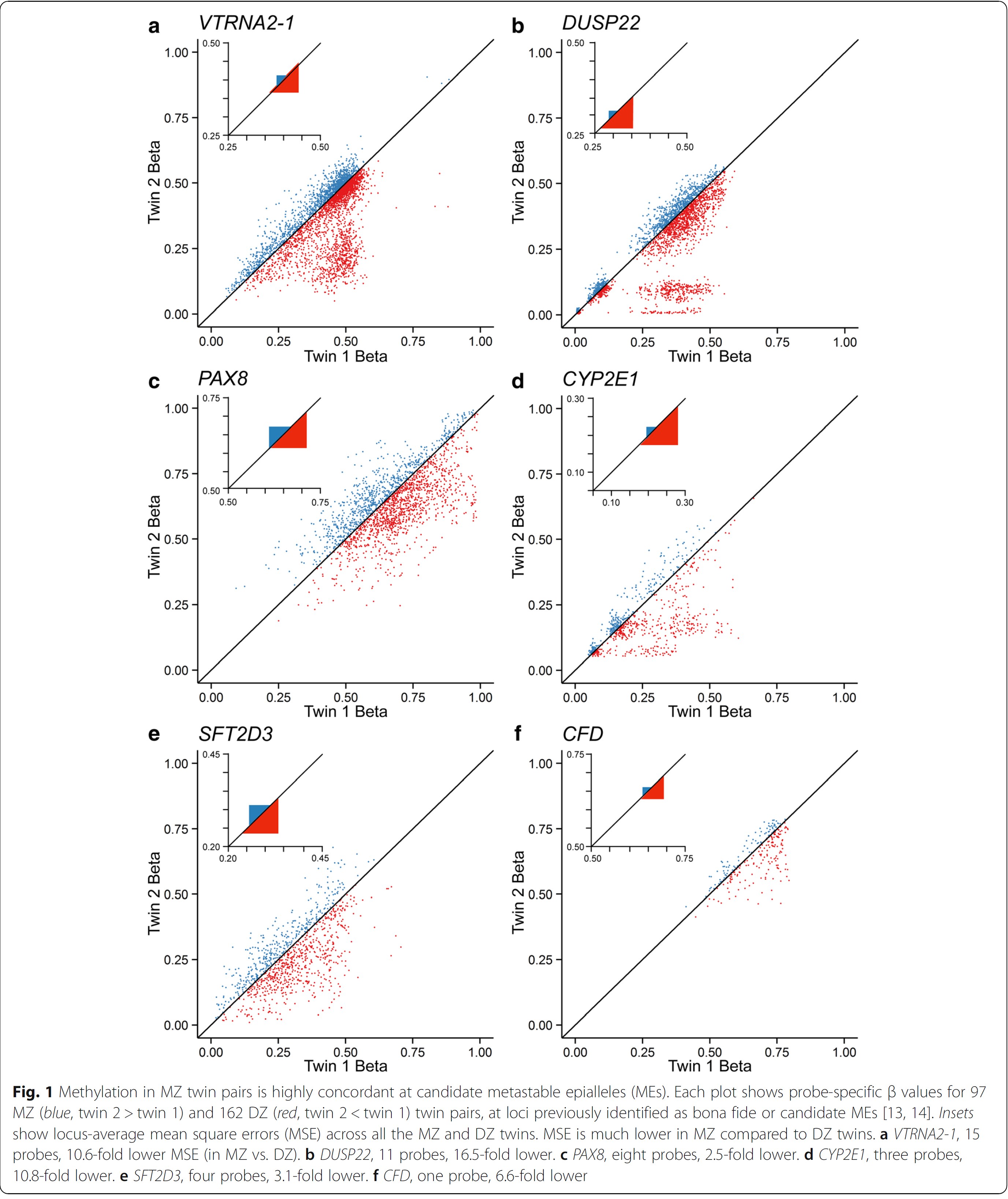
This lead van Baak et el. to look for the origin of this invariable methylation between two individuals. They found this supersimilarity between pairs of monozygotic twins  is established at or before the stage when the twins become twins. This was fairly unexpected.

During their search the authors noted that subtelomeric regions are not only important targets for methylations, but at the origin of diseases e.g. tumour later in life.

Since the paper is very technical, it is difficult to read. The fact that monozygotic twins are not only identical in the genome, but extremely similar in their epigenome, too, is worth some labour with the text.

The half-hole

The Nobelprice in Physics was awarde today to the three british researchers David Thouless, Duncan Haldane and Michael Kosterlitz for their contribution of the research on ultrathin materials, the plains of the mattter. They used a certain tool, the topology. What I didnot realize so far is that topology is the science of holes: You can not transform a hoop into a ball, the former has a hole in it, the latter not. There are structures with 0,1, 2, 3, … holes which can not be transformed into each other. Interesting.

The Nobel juror Thors Hans Hansson explained this and mentioned that holes can only be whole-numbered. There are not half-holes. This reminded me of Kurt Tucholsky, German pacifist and writer in the first part of the last century who published under the pseudonym Peter Panther a “Zur soziologischen Psychologie der Löcher“, the only English translation I found on this website: http://www.theavanwoland.com/thea_tul_text.html.

I dare to publish some lines from there:

The Philosophic Sociology of the Hole ( by Peter Panther alias Kurt Tucholsky)
A hole is there, where some-thing is not.
The hole is  the eternal companion of the non-hole.
Hole on it’s own, there is no such a thing, sorry.

If there would be something everywhere, there wouldn’t be any hole,
but there wouldn’t be any philosophy either and certainly no religion, as such that comes out of it.

The mouse couldn’t live without it, neither does man.
It’s the last chance for both of them,  when they are threatened by
the material.
Hole – is always good.

The hole is the fundament of social order and that’s how society is…

Tucholsky would have liked this Nobel prize very much.

Pdf/epub version of Hormones and the Endocrine System

Finally, all versions of the book have been approved and will shortly become available. The printed version has acquired an erratum. And in the electronic versions many mistakes and errors had to be amended. These did not change the text but the appearance of the text on the display. I might be responsible for remaining conceptual errors or for misreading articles. I would be glad if you could pass me a message when you think I was wrong.


Hormones and the Endocrine System

HOrmones and the Endocrines System
Hormones and the Endocrine System

The book has finally been published. This version is the translation of our German book 3rd edition, with some updates due to very recent new developments. The part on Juvenile Hormones was critically read by Prof. Riddiford from the USA and is thus up-to-date. I like to say thank you so much to numerous colleagues who have contributed articles that this book could be written in its present form. It has been a pleasure to work with the Springer people who produced the book. Thank you to Dr. Britta Müller and Martina Himberger who took the burden to defend and to publish this book. It has been a continious pleasure to get the help form the tex-community where I got posed many questions to make the book look nice. Among those who have contributed tremendously by reading large parts of the German and English version are my wife Beate who helped with the original version, Prof. Hubertus Jarry  in Göttingen and Prof. Ashley Grossman now in Oxford who encouraged me to go on with the English version after reading the 80 % translated and uncorrected intermediate. When you find this book enlightning, please write to me. If you think that issues are wrongly presented, please write to me that I improve the book in a next version. It has been a pleasure to prepare this book, it is an even greater pleasure to see it coming out.

Genetic immune defects at the origin of severe infectious diseases?

It seems feasible that infectious diseases have a genetic background. Most propably is a genetic defect in an immune response gene. But they have been obviously overlooked in the last 50 years. A review Severe infectious diseases of childhood as monogenic inborn errors of immunity by Jean-Laurent Casanova in PNAS demonstrates that it has not penetrated the medicinal community that inborn errors leading to infectious disease are more than normal.

Infectious Agent disease protein affected Mutation
Plasmodium vivax   Duffy antigen and receptor for chemokine (DARC) prevented disease
HIV   CCR5 prevented disease
Norovirus   fucosyl transferase 2 (FUT2) prevented disease
Mycobacterium tuberculosis Mycobacterial disease (MSMD) IFN-γ and related proteins or receptors, IL12RB1 caused disease
Neisseria Menigitis Complement C5-C9, factor D, properdin caused disease
Human Papillovirus 5 (HPV-5) Epidermodysplasia verruciformis transmembrane channel-like
6 and 8 (TMC6 and TMC8)
causing disease
Epstein-Barr virus X-linked recessive
lymphoproliferative disease (XLP)
signaling lymphocytic activation molecule-associating protein (SAP) causing disease
Candida albicans chronic mucocutaneous candidiasis (CMC) IL17F, IL-17 receptor A (IL17RA), IL17RC,  actin-related gene 1 (ACT1) causing disease
dermatophytes, Candida, Phialophora,
Exophialia, and others
Dermatophytosis (athlete’s foo) caspase recruitment domain family member
9 (CARD9)
causing disease
invasive pneumococcal disease (IPD) NF-κB essential modulator (NEMO), IL-1R–associated kinase-4 (IRAK-4),myeloid differentiation primary response gene 88 (MyD88)
 Herpex simplex herpes simplex encephalits (HSE)  UNC93B1 and thus TLR-3 causing disease
Trypanosoma evansi apolipoprotein L-I (APOL1) causing disase
Influenza virus influenza IFN regulatory factor 7 (IRF7) causing disease

The review is very suggestive. I would like to point out, that infections where no inborn error has yet been found, should not be considered to have no genomic background. The list is too impressive already to be dismissed.

Since the paper is OPEN ACCESS a must!

“Hormones and the Endocrine System” will be available soon

We are proud to announce this book to be available in the first weeks of january 2016. It has been proof-read and needs final approval. It has been quite a challenge of interactions between the Black Forest — Heidelberg — England and Chenai/Indien, but we have overcome all the difficulties which arose. In its largest part it is a translation of the German 3rd Edition of “Hormone and Hormonsystem”, but some parts about the Juvenile Hormone and its receptor have been rewritten and differ from the German original.

HOrmones and the Endocrines System
Hormones and the Endocrine System

I would like to receive comments and questions when  you happen to read the book.

A proteinaceous biomagnet?

Magnetism is helping animals to find their destinations, it does not matter whether they are very large like whales, small  like pigeon, or very tiny like butterflies  or bees, they are all dependent on the Earth magnitic field. The underlying mechanism how a magnetic field is recognized in biological terms is almost not understood.

It has been found already that the cryptochromes (Cry) have a role in magnetic reception since Cry-negative Drosophila loose their sensitivity to magnetic fields (see Gegear, R. J., Casselman, A., Waddell, S. & Reppert, S. M. Cryptochrome mediates light-dependent magnetosensitivity in Drosophila. Nature 454, 10141018 (2008)). Crytochromes are part of the intrinsic circadian clock, which resides in the human brain in the Suprachiasmatic Nucleus of the hypothalamus.

A group from Bejing has now presented a debated paper in Nature Materials that claims to have discovered a protein structure that is able to measure the magnetic field. The group around Can Xie has found that the Drosophila protein CG8198 forms complexes with FAD and Cry that establish a biomagnet. This is an astonishing piece of work. It is much debated, for example, for quantitative reasons: magnets have many more iron molecules to be seen orientating according to the field around. These tiny magnets which are in single cells, intracellular not intercellular, do not seem to have the effect necessary to communicate a message about the orientation. Whether this or the contrary will be confirmed by independent experiments, you can only guess. One could guess that cell cooperation will help to make the output from single cells large enough to become relevant. Nature there is a News & Yiews article about this paper, which is also worth reading.

This is very exciting and should be followed-up. Highly recommended!

Sleep in preindustrial societies

There are complaints that sleep in industrial communities (with electric light, noise all over) is severely compromised. These complaints have already been raised about 150 years ago, at the beginning of the industrialization. Sleep before is supposed to be calmer and longer.

This seems not to be true. Measuring the sleep durations and sleep onset and wakening in three societies that have no electric light, that remain in a pre-industrial state even in the 21st century, Yetish and colleagues report in Current Biology that sleep duration are the same as in industrial settings. Measurements were done in the last corner of Bolivia, in the Tsimane community, in Central Africa with the Ju/’hoansi San people, and in northern Tanzania with the Hadza.

There is an News and Views article on the same issue in Nature. Both articles are highly recommended.

Self-Tolerance by a second mechanism

T lymphocytes like B lymphocytes are puzzle masters that stick their receptors together in innumerable ways which is called recombination. Being free to choose from gene pieces and not concerned which receptor is finally built in a cell, it is inevitable that there are generated lymphocytes which recognize self-determinants and might attack the very same organism if not silenced from the beginning. T lymphocytes learn in the thymus to discriminate between self and non-self and only non-self reactive T cells are to leave the thymus to become eventually effector cells of the immune system, otherwise autoimmunity will develop.

The process by which any self peptide is presented to T cells has been a riddle for a long time.  From the 2000s on the riddle was partially solved: There is stochastic expression of tissue restricted selfantigens in medullary thymic epithelial cells (mTec) under the control of the AIRE protein. (See AIRE in OMIN). Stochastic means that the antigens are randomly expressed, only some in a given cell, but obviously all self antigens at a given time since autoimmunity is rare. This is achieved by an random epigenetic activation of genes. These proteins are then processed and their peptides loaded into the peptide pockets of major histocompatibility proteins class I (MHC class I). The peptides are presented in these MHC class I to the T cells which become silent once they recognize any of this peptides in self MHC. This mechamism has been about for almost 10 years.

A recent paper in Cell by Japanese colleagues adds a second mechanism to central tolerance. This time it is the Fezf2 that directs a mechanism of promiscuous gene activation. And as in the case of AIRE the lack of Fezf2 leads to autoimmune disease since autoimmune T cells are not silenced in the thymus. Fezf2 is regulated by the lymphotoxin-beta receptor. It is striking that there are genes activated by AIRE while others are stimulated by Fezf2.

After 10 years of AIRE in the air Fezf2 is a welcomed addition to the puzzle autoimmunity still presents.

Highly recommended.


Steroids, diffusion and vesicle export

Steroid hormone are believed to cross membranes by diffusion. However, Winfried Hanke of the University of Karlruhe (Hanke,W. (1970). Hormone. Fortschr Zool 20. 318–380) had evidence that corticosterone was exported in vesicles. This discussion has relevance since steroid hormones in vesicles would be released when the release is triggered and not continously, while release by diffusion would be continously and regulated by the rate-limiting enzyme in the biosynthetic pathway. And the availability of steroid hormones is one very intruiging question in endocrinology.

An arcticle in Cell of this week demonstrates that ecdysone, that steroid hormone of insects is well released in vesicles. They analyze the machinery of SNARE’s (the proteins who will eventually upon a calcium trigger fuse to the cell membrane release the vesicle content to the outside of the cell) and the loading of ecdysone to the vesicle by the Abet ATP-binding cassette transporter in detail and very convincingly.

One wonders whether other steroids are loaded into vesicles similarily. And old story is to become exciting. Nice paper and very instructive images! Highly recommended!