Category Archives: Biochemistry

A proteinaceous biomagnet?

Magnetism is helping animals to find their destinations, it does not matter whether they are very large like whales, small  like pigeon, or very tiny like butterflies  or bees, they are all dependent on the Earth magnitic field. The underlying mechanism how a magnetic field is recognized in biological terms is almost not understood.

It has been found already that the cryptochromes (Cry) have a role in magnetic reception since Cry-negative Drosophila loose their sensitivity to magnetic fields (see Gegear, R. J., Casselman, A., Waddell, S. & Reppert, S. M. Cryptochrome mediates light-dependent magnetosensitivity in Drosophila. Nature 454, 10141018 (2008)). Crytochromes are part of the intrinsic circadian clock, which resides in the human brain in the Suprachiasmatic Nucleus of the hypothalamus.

A group from Bejing has now presented a debated paper in Nature Materials that claims to have discovered a protein structure that is able to measure the magnetic field. The group around Can Xie has found that the Drosophila protein CG8198 forms complexes with FAD and Cry that establish a biomagnet. This is an astonishing piece of work. It is much debated, for example, for quantitative reasons: magnets have many more iron molecules to be seen orientating according to the field around. These tiny magnets which are in single cells, intracellular not intercellular, do not seem to have the effect necessary to communicate a message about the orientation. Whether this or the contrary will be confirmed by independent experiments, you can only guess. One could guess that cell cooperation will help to make the output from single cells large enough to become relevant. Nature there is a News & Yiews article about this paper, which is also worth reading.

This is very exciting and should be followed-up. Highly recommended!
Steroids, diffusion and vesicle export

Steroid hormone are believed to cross membranes by diffusion. However, Winfried Hanke of the University of Karlruhe (Hanke,W. (1970). Hormone. Fortschr Zool 20. 318–380) had evidence that corticosterone was exported in vesicles. This discussion has relevance since steroid hormones in vesicles would be released when the release is triggered and not continously, while release by diffusion would be continously and regulated by the rate-limiting enzyme in the biosynthetic pathway. And the availability of steroid hormones is one very intruiging question in endocrinology.

An arcticle in Cell of this week demonstrates that ecdysone, that steroid hormone of insects is well released in vesicles. They analyze the machinery of SNARE’s (the proteins who will eventually upon a calcium trigger fuse to the cell membrane release the vesicle content to the outside of the cell) and the loading of ecdysone to the vesicle by the Abet ATP-binding cassette transporter in detail and very convincingly.

One wonders whether other steroids are loaded into vesicles similarily. And old story is to become exciting. Nice paper and very instructive images! Highly recommended!

RNA polymerase in action

To see an RNA polymerase II (Pol II) in action has been the dream of any molecular biologist. Imaging the possibilities to see and not to suspect transcription of DNA into RNA, to see what the interactions are and not to suppose. A team around Guillermo Calero in Pittsburgh, PA, and Craig D. Kaplan at Texas, A & M University, has achieved this molecular dream.

The paper in Molecular Cell by C.O. Barnes and M. Calero depicts the crystal structure of a RNA Pol II complex together with DNA and the newly transcribed RNA stabilized by transcription factor II F (TFIIF). The structure from Saccharomyces cerevisiae reveals  the unwinding of the the DNA duplex, the so-called nucleic acid scaffold (NAS) where the RNA is formed and the downstream duplex. The down stream duplex builds a 130 ° degree angle to the upstream duplex in the complex.  The pictures show for the first time the unwinding of DNA, the RNA synthesis, the re-winding, and the contributions of the different Pol II subunits.

Unfortunately the accession numbers for the structure are not yet availabe in the Protein Data Bank (5C4X/A/4/J, 5C3E). (They are now.)

This paper is a must for any one teaching molecular biology, for students in any case.


New Malaria drug with DDD107498

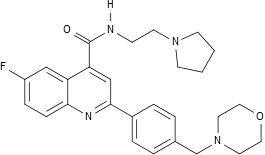
200 millions of cases and more than 500000 death are worth every effort to deal with the world wide pest named malaria. A report in nature from a multicentric group with the Welcome Trust and Glaxo has found a new drug with the so-far non-attractive name DDD107498, which has excellent non-clinical parameters. It is drug specifically active against the translation elongation factor 2 (eEF2) of Plasmodium falciparum . 

DDD107498

A patent for the drug is filed (PCT/GB2009/002084).

Wrong splicing of the androgen receptor as the cause for polycystic ovary syndrome (?)

It has since long been suggested that androgen receptor changes are at the origin of the polycystic ovary syndrome (PCO), which affects about 7 % of fertile women and is a major cause for infertility; good proof, however, has been lacking. Wang and colleagues from the Hangzhou University in China have now presented in PNAS from April 15 this year a convincing report that alternative splice variants (ASV) occur in women with PCO but not in those without. 

Alternative splicing occurs when there are several acceptor sites for the RNA lariat during splicing, where the introns are excised from the heteronuclear RNA and the RNA is cut to the messenger RNA. Or there are mutations at the sites supposed to be brought together that the splicing mechanism can no longer work.

It is very suggestive that the ASV occuring in the androgen receptor are causative for the disease. It is very much supprising that this finding has taken so much time to be discovered. This lets one think about lack of basic scientific knowlege in the medical community at large. It should be necessary to have scientists advisors assisting medical researchers not beeing able to look beyond their own nose.

An important paper and a must.

Prolactin acts on stem cells – understanding circannual behaviour?

Prolactin is the hormone that regulates mammary gland development in man. However, in animals it is the main functional regulator to transmit the physiological reaction to the seasons, its expression is dependent on the day length, which is measured in calendar cells close to the hypophyseal stalk, and which activate prolactin expression when the day time increases, and vice versa.

How prolactin could in turn influence different functions such as increase in mating behaviour, coat colour changes or molt, the song in birds, e.g., has been an open question. A minireview in Molecular Endocrinology by Sackmann-Sala and colleagues from the  Institut Necker in Paris, France, may shed light on this issue. They show that in humans, mice, and rats prolactin acts on stem cells in a tissue specific way. The tissues in question are reproductive tissues, but apart from that also special regions of the brain, and peripheral tissues.

If each of the functions as mediated from the progeny of individual stem cells, then a stimulating role of the pleiotropic prolactin activities is easily understood. The paper does not address this question, but it opens a new way of thinking.

For this reason, do not miss it if you are concerned with circannual regulation. 

Not seeing what is obvious

Sometimes we need an eye-opener to notice things which are in our reach and still beyond comprehesion. We smite our forehead and confess how could we be such blinds. The paper “The essence of female–male physiological dimorphism: Differential Ca2+-homeostasis enabled by the interplay between farnesol-like endogenous sesquiterpenoids and sex-steroids? The Calcigender paradigm” by Arnold de Loof in “General and Comparative Endocrinology” is such an eye-opener. It puts the Calcium (Ca) metabolism in an entire new prospective. De Loof argues that Ca is a toxin which has to be kept away and that reproductive activities such as egg laying and mild production have developed for this purpose since the Ca that an egg contains and which is in milk deplete the organism of much Ca.

The Ca concentration in blood is in the range of 1-3 mM in man and are conserved in evolution. In the cell however, there are only 100 nM, ten times less. The cell membrane is partially permeable for Ca so that the cell always has to deal with an excess of Ca which is permanently depleted by Ca export or/and storage in entoplasmatic reticulum vesicles.

The surprise of the paper is that de Loof finds a function for farnesol in eukaryotes: A Ca transporter in the ER that is only regulated by farnesol-like substances is common to vertebrates and insects.

Whether all the ideas of the paper will survive experimental scrutiny is to be seen, but two ideas stick: Calcium as a toxin and its role or more to the point the removal of Calcium at the origin of reproductional activity.

The paper is open access and and a must.

Sirtuin protein at the start of GnRH neuron migration

The GnRH neurons are unique among the hypothalamic neurons that they originate not in hypothalamus itself, but in the vomeronasal organ of the olfactory bulb and move (in the mouse) between day 10 and 17 of embryonic development into the hypothalamus via the forebrain. When this wandering is impaired, there is not any GnRH synthesis in the hypothalamus due to missing GnRH neurons, a phenomen called Kallmann syndrome, and subsequently the patient undergo hypogonadotrophic hypogonadism.

In a report in Molecular Endocrinology this week Gabriel Di Sante and colleagues from Philadelphia with the help of Canadian coworkers from Ottawa describe in mice another protein involved in this wandering of neurons. They found that the Sirt1 protein is necessary to start the migration of GnRH neurons. Sirt1 is the analogue of sirtuin protein originally found in yeast as Silent regulatory protein and has diverse physiological functions. Sirt1 defective mutants are not viable and die in utero.

The paper shows that the migration is initiated intracellularly due to the interaction of FGF8 and the FGF receptor, Sirt1, and corstatin, whereupon the sirtuin protein leaves the nucleus and deacylates the cytoplasmatically located corstatin. This interaction then makes the neuron migrate. There are other mechanisms listed in the introduction of the article which effect the migration. But none is as near to the origin of the migration as this one.

A nice piece of work! Recommended!

Seeing that they see differently

What we see is determined by the visual pigments of the human eye. We have often asked what our dog might see. Colour vision was thought restricted to the primates. Totally wrong. Thanks to a paper by Justin Marshall & Kentaro Arikawa in Current Biology we know now that colour vision is very common in the animal kingdom. There are several species that use a much broader spectrum to look at their surroundings. Honey bees for example can identify objects in the UV part of the spectrum. They have, however, three different photoreceptors like humans. Waterflees or the Blue Tit have four different ones the former sensing the whole spectrum from 300 nm to 700 nm. The latter misses some light in the infrared part. Horses and dogs have only two different photoreceptors, they are in way green–red blind. Dogs, however, have a much more discriminating capacity in the dark.

What comes as a surprise is that butterflies and other insects can differentiate the visual reception with up to 8 different photoreceptors. And this is not the end: Shrimps have 20: (cited from the paper)”Twenty receptor types have been defined: twelve for colour, six for polarisation and two with overlapping function for luminance tasks”.

An eye-opener. Nice and Recommende!