Category Archives: Cell physiology

A proteinaceous biomagnet?

Magnetism is helping animals to find their destinations, it does not matter whether they are very large like whales, small  like pigeon, or very tiny like butterflies  or bees, they are all dependent on the Earth magnitic field. The underlying mechanism how a magnetic field is recognized in biological terms is almost not understood.

It has been found already that the cryptochromes (Cry) have a role in magnetic reception since Cry-negative Drosophila loose their sensitivity to magnetic fields (see Gegear, R. J., Casselman, A., Waddell, S. & Reppert, S. M. Cryptochrome mediates light-dependent magnetosensitivity in Drosophila. Nature 454, 10141018 (2008)). Crytochromes are part of the intrinsic circadian clock, which resides in the human brain in the Suprachiasmatic Nucleus of the hypothalamus.

A group from Bejing has now presented a debated paper in Nature Materials that claims to have discovered a protein structure that is able to measure the magnetic field. The group around Can Xie has found that the Drosophila protein CG8198 forms complexes with FAD and Cry that establish a biomagnet. This is an astonishing piece of work. It is much debated, for example, for quantitative reasons: magnets have many more iron molecules to be seen orientating according to the field around. These tiny magnets which are in single cells, intracellular not intercellular, do not seem to have the effect necessary to communicate a message about the orientation. Whether this or the contrary will be confirmed by independent experiments, you can only guess. One could guess that cell cooperation will help to make the output from single cells large enough to become relevant. Nature there is a News & Yiews article about this paper, which is also worth reading.

This is very exciting and should be followed-up. Highly recommended!
Sleep in preindustrial societies

There are complaints that sleep in industrial communities (with electric light, noise all over) is severely compromised. These complaints have already been raised about 150 years ago, at the beginning of the industrialization. Sleep before is supposed to be calmer and longer.

This seems not to be true. Measuring the sleep durations and sleep onset and wakening in three societies that have no electric light, that remain in a pre-industrial state even in the 21st century, Yetish and colleagues report in Current Biology that sleep duration are the same as in industrial settings. Measurements were done in the last corner of Bolivia, in the Tsimane community, in Central Africa with the Ju/’hoansi San people, and in northern Tanzania with the Hadza.

There is an News and Views article on the same issue in Nature. Both articles are highly recommended.

Steroids, diffusion and vesicle export

Steroid hormone are believed to cross membranes by diffusion. However, Winfried Hanke of the University of Karlruhe (Hanke,W. (1970). Hormone. Fortschr Zool 20. 318–380) had evidence that corticosterone was exported in vesicles. This discussion has relevance since steroid hormones in vesicles would be released when the release is triggered and not continously, while release by diffusion would be continously and regulated by the rate-limiting enzyme in the biosynthetic pathway. And the availability of steroid hormones is one very intruiging question in endocrinology.

An arcticle in Cell of this week demonstrates that ecdysone, that steroid hormone of insects is well released in vesicles. They analyze the machinery of SNARE’s (the proteins who will eventually upon a calcium trigger fuse to the cell membrane release the vesicle content to the outside of the cell) and the loading of ecdysone to the vesicle by the Abet ATP-binding cassette transporter in detail and very convincingly.

One wonders whether other steroids are loaded into vesicles similarily. And old story is to become exciting. Nice paper and very instructive images! Highly recommended!

RNA polymerase in action

To see an RNA polymerase II (Pol II) in action has been the dream of any molecular biologist. Imaging the possibilities to see and not to suspect transcription of DNA into RNA, to see what the interactions are and not to suppose. A team around Guillermo Calero in Pittsburgh, PA, and Craig D. Kaplan at Texas, A & M University, has achieved this molecular dream.

The paper in Molecular Cell by C.O. Barnes and M. Calero depicts the crystal structure of a RNA Pol II complex together with DNA and the newly transcribed RNA stabilized by transcription factor II F (TFIIF). The structure from Saccharomyces cerevisiae reveals  the unwinding of the the DNA duplex, the so-called nucleic acid scaffold (NAS) where the RNA is formed and the downstream duplex. The down stream duplex builds a 130 ° degree angle to the upstream duplex in the complex.  The pictures show for the first time the unwinding of DNA, the RNA synthesis, the re-winding, and the contributions of the different Pol II subunits.

Unfortunately the accession numbers for the structure are not yet availabe in the Protein Data Bank (5C4X/A/4/J, 5C3E). (They are now.)

This paper is a must for any one teaching molecular biology, for students in any case.


Puberty – new evidence for its regulation

It has been a long standing mystery how the start of puberty is initiated. Fact is that menarche, the begin of active reproduction capacity is preceded by and a consequence of the adrenarche, the begin of androgen production in the adrenal gland. Puberty is characterized by the beginning of sporadic gonadotropin releasing hormone (GnRH) pulses which in time become regular and finally acquire their one-in-two hour rhythm at the end of puberty.

A report in the Journal of Molecular Endocrinology by Abreu and colleagues from Boston and Sao Paulo has uncovered  that the Makorin ring finger 3 (MKRN3) gene is mutated in cases of central precocious puberty (CPP) . CPP is diagnosed when the children enter into puberty much to early for their age. They analyzed the protein in more detail then and found the decline of MKRN3 expression in the arcuate nucleus (area of the hypothalamus to control GnRH secretion) is necessary for the increase of GnRH secretion. Without GnRH puberty can not take place. By which stimulus the decline of MKRN3 is initiated has not been described. It is discussed whether MKRN3 acts directly on GnRH secretion or on kisspeptin, neuromedin B or dynorphin, known mediators of GnRH secretion. It can not act on GnRH expression since the GnRH neurons only reach with their axons into the arcuate nucleus where their release is controlled by other neurons and mediators.

This is a nice paper, adding valuable information to people concerned with the mechanisms of puberty. Recommended!

Wrong splicing of the androgen receptor as the cause for polycystic ovary syndrome (?)

It has since long been suggested that androgen receptor changes are at the origin of the polycystic ovary syndrome (PCO), which affects about 7 % of fertile women and is a major cause for infertility; good proof, however, has been lacking. Wang and colleagues from the Hangzhou University in China have now presented in PNAS from April 15 this year a convincing report that alternative splice variants (ASV) occur in women with PCO but not in those without. 

Alternative splicing occurs when there are several acceptor sites for the RNA lariat during splicing, where the introns are excised from the heteronuclear RNA and the RNA is cut to the messenger RNA. Or there are mutations at the sites supposed to be brought together that the splicing mechanism can no longer work.

It is very suggestive that the ASV occuring in the androgen receptor are causative for the disease. It is very much supprising that this finding has taken so much time to be discovered. This lets one think about lack of basic scientific knowlege in the medical community at large. It should be necessary to have scientists advisors assisting medical researchers not beeing able to look beyond their own nose.

An important paper and a must.

Regulatory T cells and AIRE expressing cells do the job of tolerance

T lymphocytes require education in order to distinguish between self and nonself. This education is maintained by thymus stromal epithelial expressing the autoimmune regulator gene (AIRE). These cells express proteins from throughout the organism not in promotor regulated way, but in a epigenetically controlled statistically mode, the only cells that do so. This has as a consequence that these cells present on their surface each a part of the possible plethora of self peptides in the context of MHC class I molecule. T cells which react with them do recognize self and should be suppressed lifelong to avoid autoimmunity.

In a paper by Yang and colleagues from the Benoist/Mathis laboratory at Stanford it is now reported, that early regulatory T cells are additionally required to maintain the protection against autoimmune diseases. If the cells are lacking due to the absece of AIRE expression, a disease called autoimmune polyendocrinopathy–candidiasis–ectodermal dystrophy or
autoimmune polyglandular syndrome 1 is the consequence. The News and Views contribution by Tanaka and Sakaguchi explains this in detail.  This is a nice addition to the problem of tolerance and and might be an important step to development of tolerance to foreign antigens as well. That would be required to make gene therapy sustainable.

A must!

Prolactin acts on stem cells – understanding circannual behaviour?

Prolactin is the hormone that regulates mammary gland development in man. However, in animals it is the main functional regulator to transmit the physiological reaction to the seasons, its expression is dependent on the day length, which is measured in calendar cells close to the hypophyseal stalk, and which activate prolactin expression when the day time increases, and vice versa.

How prolactin could in turn influence different functions such as increase in mating behaviour, coat colour changes or molt, the song in birds, e.g., has been an open question. A minireview in Molecular Endocrinology by Sackmann-Sala and colleagues from the  Institut Necker in Paris, France, may shed light on this issue. They show that in humans, mice, and rats prolactin acts on stem cells in a tissue specific way. The tissues in question are reproductive tissues, but apart from that also special regions of the brain, and peripheral tissues.

If each of the functions as mediated from the progeny of individual stem cells, then a stimulating role of the pleiotropic prolactin activities is easily understood. The paper does not address this question, but it opens a new way of thinking.

For this reason, do not miss it if you are concerned with circannual regulation. 

Angiotensin-Receptor in 3D

Almost all the physiological actions of angiotensin II, the effective mediator after renal renin has cleaved the precursor angiotensinogen and the angiotensin-converting enzyme (ACE) of the lung has liberated angiotensin II from angiotensin I, are mediated by by the angiotensin II type 1 receptor. It is a G-protein coupled receptor (GPcR) like many hormone receptors of the rhodopsin family (Omin 106165). Like many GPcR it has been difficult to crystallize to dertermine its threedimensional structure.

Zhang and colleagues from Los Angeles and other places have now used femtosecond chrystallography to reveal the structure with the high-affinity agonist ZD71DD as ligand.

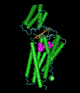
AT1R
AT1R (image with the free tool Cn3D from NIH and the sequence 4YAY from MMDB)

The depicted image is nothing compared to the images in the paper. You can, however, see how the ligand fits in a binding pocket in the transmembrane domain with its numerous helices. The domain on top is a extracellular domain.

The structure should help to resolve questions concerning the regulation of blood pressure, how mutations influence the binding of angiotensin II and may help to develop other drugs.

This is a very nice piece of work. Highly recommended!

Melatonin required to fall asleep – in fish

Reading about circadian rhymth in flies I happened to see the Neuron paper by Gandhi et al. about melatonins role in fish. Melatonin – the hormone of the pineal gland – has been shown already to be active in the determination of seasons, its amount produced during the night being measure in so-called calendar cells in the vicinity of the hyphyseal stalk. Now the authors in Pasadena show that melatonin is necessary to fall asleep: zebrafish without the critical enzyme of melatonin systhesis: aanat2 (arylalkylamine N-acetyltransferase 2) take much longer to fall asleep and do not sleep as long as control animals.

Whether the data do apply to men and mammals is open. This is, however, a nice piece of work. It does not explain while I can start sleeping extensively during day time when there is not any melatonin in my circulation.