Category Archives: Hormones

Puberty – new evidence for its regulation

It has been a long standing mystery how the start of puberty is initiated. Fact is that menarche, the begin of active reproduction capacity is preceded by and a consequence of the adrenarche, the begin of androgen production in the adrenal gland. Puberty is characterized by the beginning of sporadic gonadotropin releasing hormone (GnRH) pulses which in time become regular and finally acquire their one-in-two hour rhythm at the end of puberty.

A report in the Journal of Molecular Endocrinology by Abreu and colleagues from Boston and Sao Paulo has uncovered  that the Makorin ring finger 3 (MKRN3) gene is mutated in cases of central precocious puberty (CPP) . CPP is diagnosed when the children enter into puberty much to early for their age. They analyzed the protein in more detail then and found the decline of MKRN3 expression in the arcuate nucleus (area of the hypothalamus to control GnRH secretion) is necessary for the increase of GnRH secretion. Without GnRH puberty can not take place. By which stimulus the decline of MKRN3 is initiated has not been described. It is discussed whether MKRN3 acts directly on GnRH secretion or on kisspeptin, neuromedin B or dynorphin, known mediators of GnRH secretion. It can not act on GnRH expression since the GnRH neurons only reach with their axons into the arcuate nucleus where their release is controlled by other neurons and mediators.

This is a nice paper, adding valuable information to people concerned with the mechanisms of puberty. Recommended!
Wrong splicing of the androgen receptor as the cause for polycystic ovary syndrome (?)

It has since long been suggested that androgen receptor changes are at the origin of the polycystic ovary syndrome (PCO), which affects about 7 % of fertile women and is a major cause for infertility; good proof, however, has been lacking. Wang and colleagues from the Hangzhou University in China have now presented in PNAS from April 15 this year a convincing report that alternative splice variants (ASV) occur in women with PCO but not in those without. 

Alternative splicing occurs when there are several acceptor sites for the RNA lariat during splicing, where the introns are excised from the heteronuclear RNA and the RNA is cut to the messenger RNA. Or there are mutations at the sites supposed to be brought together that the splicing mechanism can no longer work.

It is very suggestive that the ASV occuring in the androgen receptor are causative for the disease. It is very much supprising that this finding has taken so much time to be discovered. This lets one think about lack of basic scientific knowlege in the medical community at large. It should be necessary to have scientists advisors assisting medical researchers not beeing able to look beyond their own nose.

An important paper and a must.

Man and dog – bonding and evolution

Science from April 17, this year, has two feature articles by David Grimm about men and his dog, one perspective article and a report that shows how men-dog but not men-wolf bonding is intensifierd by oxytocin.

One feature article: Dawn of the dog is most instructive about the process in science itself, fighting over data, sparring with opponents, and reconsiliation to a common task with the help of a third party, the other one: How the wolf became a dog is summarizing the different text, the perspective paper by MacLean and Hare shows the implications of the report: How the oxytozin loop between men and dog might shape the bonding and even help in pathological situation like autism and posttraumatic stress. A nice sentence from the feature is “if the dog is staring at you, it might not be after your sandwich!”

The whole bunch of articles is a pleasue to read.

Prolactin acts on stem cells – understanding circannual behaviour?

Prolactin is the hormone that regulates mammary gland development in man. However, in animals it is the main functional regulator to transmit the physiological reaction to the seasons, its expression is dependent on the day length, which is measured in calendar cells close to the hypophyseal stalk, and which activate prolactin expression when the day time increases, and vice versa.

How prolactin could in turn influence different functions such as increase in mating behaviour, coat colour changes or molt, the song in birds, e.g., has been an open question. A minireview in Molecular Endocrinology by Sackmann-Sala and colleagues from the  Institut Necker in Paris, France, may shed light on this issue. They show that in humans, mice, and rats prolactin acts on stem cells in a tissue specific way. The tissues in question are reproductive tissues, but apart from that also special regions of the brain, and peripheral tissues.

If each of the functions as mediated from the progeny of individual stem cells, then a stimulating role of the pleiotropic prolactin activities is easily understood. The paper does not address this question, but it opens a new way of thinking.

For this reason, do not miss it if you are concerned with circannual regulation. 

Parathyroid hormone essential for gill formation in zebrafish

A paper by Kwong and Perry from Ottawa in Endocrinlogy reveals that parathyroid hormone 1 (PTH1) in addition to its effects on bone formation is also necessary for the development of proper gills. This is a nice addition to the functions of PTH1 as a regulator of calcium availability.

The paper demonstrates convincingly this new role for PTH1. Recommended!

Angiotensin-Receptor in 3D

Almost all the physiological actions of angiotensin II, the effective mediator after renal renin has cleaved the precursor angiotensinogen and the angiotensin-converting enzyme (ACE) of the lung has liberated angiotensin II from angiotensin I, are mediated by by the angiotensin II type 1 receptor. It is a G-protein coupled receptor (GPcR) like many hormone receptors of the rhodopsin family (Omin 106165). Like many GPcR it has been difficult to crystallize to dertermine its threedimensional structure.

Zhang and colleagues from Los Angeles and other places have now used femtosecond chrystallography to reveal the structure with the high-affinity agonist ZD71DD as ligand.

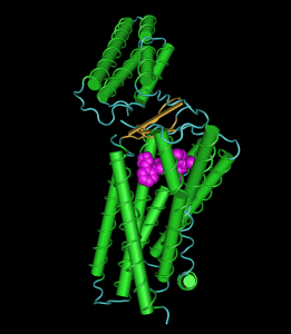
AT1R
AT1R (image with the free tool Cn3D from NIH and the sequence 4YAY from MMDB)

The depicted image is nothing compared to the images in the paper. You can, however, see how the ligand fits in a binding pocket in the transmembrane domain with its numerous helices. The domain on top is a extracellular domain.

The structure should help to resolve questions concerning the regulation of blood pressure, how mutations influence the binding of angiotensin II and may help to develop other drugs.

This is a very nice piece of work. Highly recommended!

Melatonin required to fall asleep – in fish

Reading about circadian rhymth in flies I happened to see the Neuron paper by Gandhi et al. about melatonins role in fish. Melatonin – the hormone of the pineal gland – has been shown already to be active in the determination of seasons, its amount produced during the night being measure in so-called calendar cells in the vicinity of the hyphyseal stalk. Now the authors in Pasadena show that melatonin is necessary to fall asleep: zebrafish without the critical enzyme of melatonin systhesis: aanat2 (arylalkylamine N-acetyltransferase 2) take much longer to fall asleep and do not sleep as long as control animals.

Whether the data do apply to men and mammals is open. This is, however, a nice piece of work. It does not explain while I can start sleeping extensively during day time when there is not any melatonin in my circulation.

Estrogen in insects: again — No Nein njet non

Another paper in Gen.Comp.Endo. addresses the issue “Vertebrate estrogen regulates the development of female characteristics in silkworm, Bombyx mori”. While they use vertebrate estradiol to see its effect on vitellogenin expression, they also analyse whether endogenous estrogen is present. They claim that an endogenous estrogen analogue exists in B. mori.

Very strange, to be polite. Or bullshit, to be honest. The silkworm genome has been almost fully sequenced. The side-chain cleavage enzyme (cyp11a1) are definitely not present as well as the aromatase (cyp19). Without these no chance for any estradiol or analogue.

What they may have found is an estrogen receptor agonist. The literature is full of these. They are usually called endocrine disruptors since they disturb normal estrogen functions. The structures of estrogen receptor agonists are diverse and no clearcut picture has emerged to my knowledge. Such a molecule may well be present in Bombyx mori.

Caution! Not always when estrogen is claimed there is estradiol present!

Not seeing what is obvious

Sometimes we need an eye-opener to notice things which are in our reach and still beyond comprehesion. We smite our forehead and confess how could we be such blinds. The paper “The essence of female–male physiological dimorphism: Differential Ca2+-homeostasis enabled by the interplay between farnesol-like endogenous sesquiterpenoids and sex-steroids? The Calcigender paradigm” by Arnold de Loof in “General and Comparative Endocrinology” is such an eye-opener. It puts the Calcium (Ca) metabolism in an entire new prospective. De Loof argues that Ca is a toxin which has to be kept away and that reproductive activities such as egg laying and mild production have developed for this purpose since the Ca that an egg contains and which is in milk deplete the organism of much Ca.

The Ca concentration in blood is in the range of 1-3 mM in man and are conserved in evolution. In the cell however, there are only 100 nM, ten times less. The cell membrane is partially permeable for Ca so that the cell always has to deal with an excess of Ca which is permanently depleted by Ca export or/and storage in entoplasmatic reticulum vesicles.

The surprise of the paper is that de Loof finds a function for farnesol in eukaryotes: A Ca transporter in the ER that is only regulated by farnesol-like substances is common to vertebrates and insects.

Whether all the ideas of the paper will survive experimental scrutiny is to be seen, but two ideas stick: Calcium as a toxin and its role or more to the point the removal of Calcium at the origin of reproductional activity.

The paper is open access and and a must.

Sirtuin protein at the start of GnRH neuron migration

The GnRH neurons are unique among the hypothalamic neurons that they originate not in hypothalamus itself, but in the vomeronasal organ of the olfactory bulb and move (in the mouse) between day 10 and 17 of embryonic development into the hypothalamus via the forebrain. When this wandering is impaired, there is not any GnRH synthesis in the hypothalamus due to missing GnRH neurons, a phenomen called Kallmann syndrome, and subsequently the patient undergo hypogonadotrophic hypogonadism.

In a report in Molecular Endocrinology this week Gabriel Di Sante and colleagues from Philadelphia with the help of Canadian coworkers from Ottawa describe in mice another protein involved in this wandering of neurons. They found that the Sirt1 protein is necessary to start the migration of GnRH neurons. Sirt1 is the analogue of sirtuin protein originally found in yeast as Silent regulatory protein and has diverse physiological functions. Sirt1 defective mutants are not viable and die in utero.

The paper shows that the migration is initiated intracellularly due to the interaction of FGF8 and the FGF receptor, Sirt1, and corstatin, whereupon the sirtuin protein leaves the nucleus and deacylates the cytoplasmatically located corstatin. This interaction then makes the neuron migrate. There are other mechanisms listed in the introduction of the article which effect the migration. But none is as near to the origin of the migration as this one.

A nice piece of work! Recommended!